Research projects – Group M. Nguyen Chi
AXIS 1 : VISUALIZATION OF THE DYNAMICS OF MACROPHAGE POLARIZATION
Previously we showed that zebrafish macrophages are able to switch from a pro-inflammatory phenotype to a non-inflammatory phenotype locally and in vivo. We investigate if and how macrophages switch phenotypes during wound healing using original tools developed in the lab.
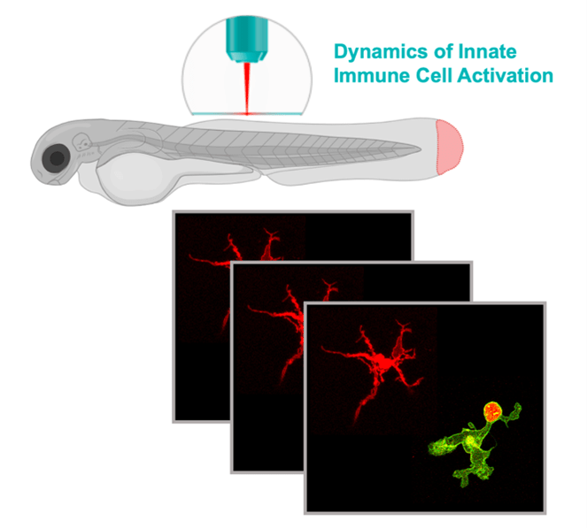
Figure 1: Combining high resolution imaging (spinning disk microscopy, SPIM) and fluorescent reporters, we are imaging in live the polarization state of macrophages.
AXIS 2 : MOLECULAR MECHANISMS UNDERLYING MACROPHAGE POLARIZATION DURING WOUND HEALING
Many signals are thought to be released in response to tissue damage. While most of these signals have been identified in vitro, it is unclear what is their real contribution in macrophage polarization in vivo. We previously showed that early wound signals (Reactive Oxygen Species and calcium) are crucial for macrophage polarization. We are now investigating the role of selected signals in macrophage polarization during wound healing.
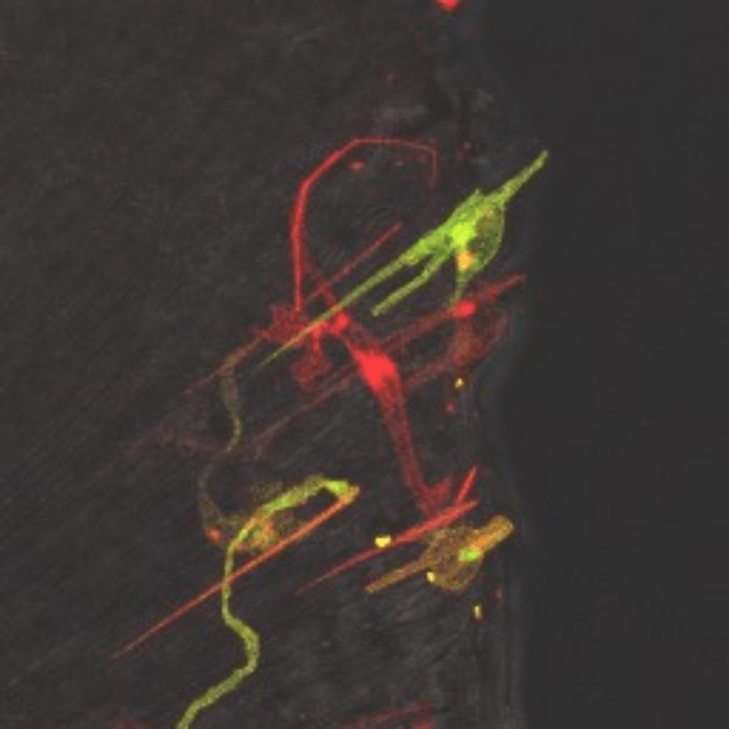
Figure 2: Macrophages recruitment to the wound following caudal fin transection in zebrafish larvae. Transgenic reporters allow the visualization of activated macrophages using different fluorescent proteins using confocale microscopy.
AXIS 3 : INTERACTION BETWEEN POLARIZED MACROPHAGES AND BACTERIA
Pathogens bacteria have evolved to survive inside their host by manipulating host defenses. Subversion of macrophage polarization has been proposed to be an important pathogenesis mechanism of intracellular bacteria. Using live imaging, we study the interactions between polarized macrophages and bacteria in a living host.
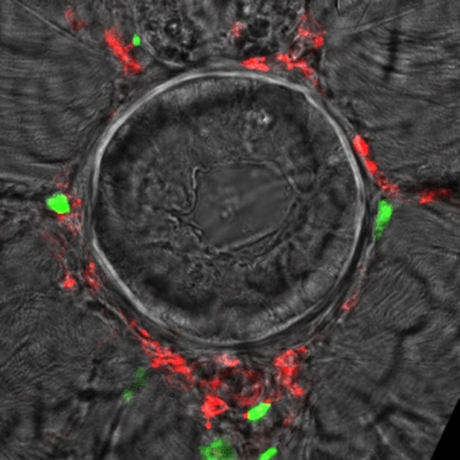
Figure 3: Recruitment of macrophages (red) and neutrophils (green) around the notochord of zebrafish larvae following infection with Escherichia coli. The role of different populations of phagocytes was studied using fluorescent reporter lines. Sectional view of the notochord.
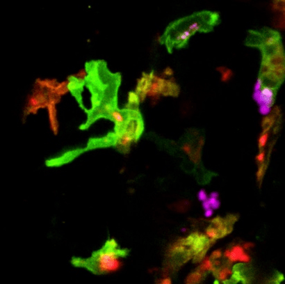
Figure 4: Interaction between polarized macrophages (tnfa- positive macrophages, red and green) and bacteria (E. coli, magenta). Confocal microscopy.