Research projects – Group A. Blanc-Potard
We aim to provide a better understanding of an intramacrophage phase for extracellular pathogens, especially in the cystic fibrosis context, as well as identify novel antimicrobial molecules targeting factors involved in intramacrophage survival.
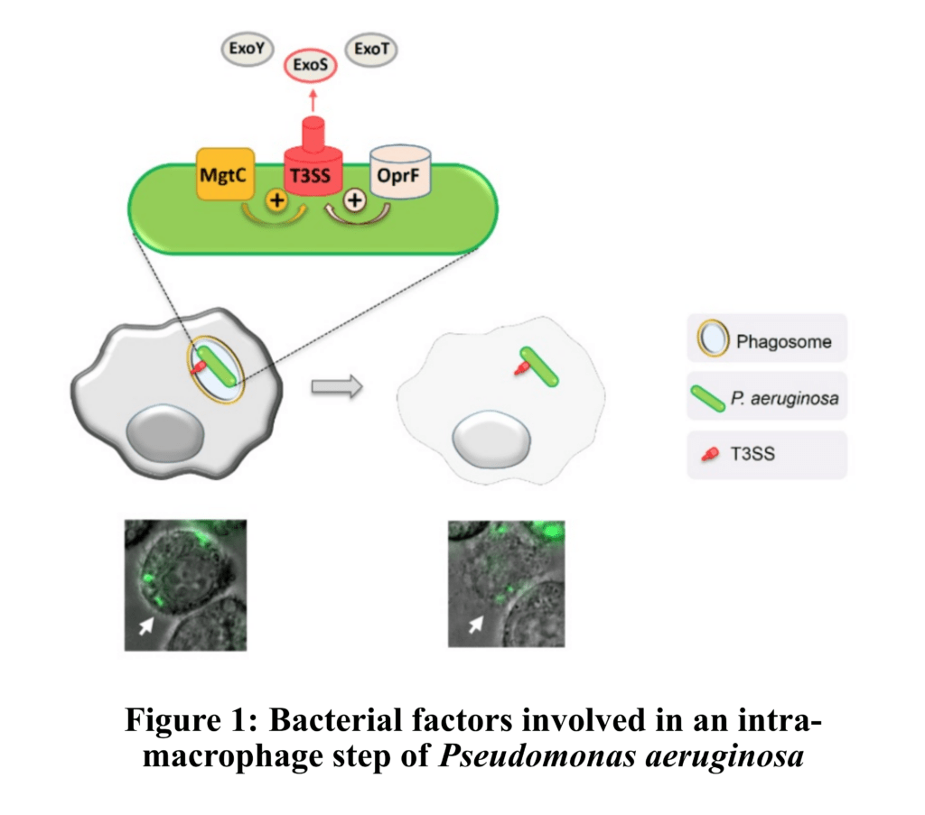
AXIS 1 : BACTERIAL FACTORS INVOLVED IN INTRAMACROPHAGE SURVIVAL
Intracellular bacterial pathogens, as Mycobacterium tuberculosis or Salmonella enterica, can replicate within phagocytic cells. In contrast, extracellular pathogens, as Staphylococcus aureus or Pseudomonas aeruginosa, avoid phagocytosis, thus promoting extracellular multiplication. However, a phase of intracellular residence can be of importance for so-called extracellular pathogens. Focusing mainly on P. aeruginosa, our objective is to investigate the function and expression of specific bacterial virulence factors (MgtC, OprF, T3SS, ExoS ..) involved in an intramacrophage stage. We then aim to decipher the role if this intracellular phase in the establishment, dissemination and persistence of the infection, using zebrafish embryos, which offers powerful tools to study host-pathogens interactions. Embryos deficient for CFTR channel are used as surrogate for cystic fibrosis context (Figure 1).
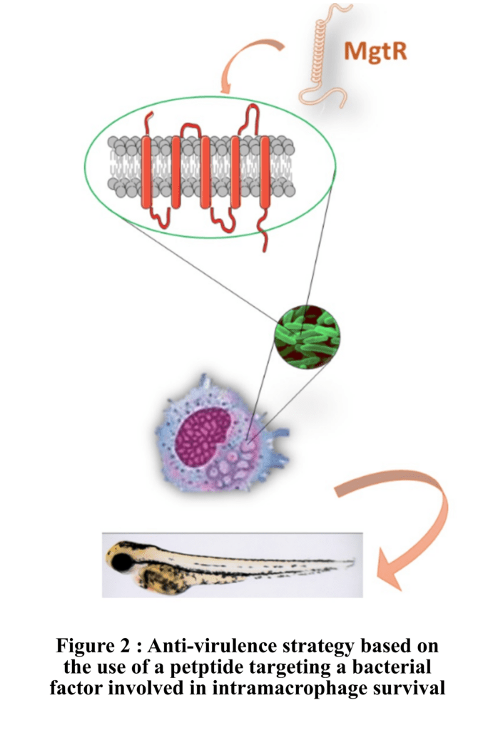
AXIS 2 : ANTI-VIRULENCE STRATEGIES
Morevover, uprising antibiotic resistance in various bacteria urges for the development of novel antibacterial strategies. Our studies of factors involved in intramacrophage survival have provided new targets for antivirulence strategies. We have recently identified hydrophobic peptides that reduce bacterial intramacrophage survival upon overexpression or exogenous addition. Our current goal is to decipher the mechanisms of action of synthetic peptides of interest and to test their antivirulence activity against various bacteria and in the zebrafish infection model. Through collaborations, we also test the efficiency of molecules inhibiting other specific targets (as NADK, T3SS ..) against P. aeruginosa using macrophage infection model and zebrafish infection model (Figure 2).